RESEARCH OVERVIEW
Precise organization of neuronal connections is essential for processing information. A major challenge in neuroscience is to understand how these connections are properly established, and how errors in this wiring process can lead to disorders. During develop-ment, neurons extend axons that navigate along defined paths towards their target by responding to attractive and repulsive cues. Concomitantly or subsequently to this guidance process, refinement mechanisms involving degeneration or pruning correct axons that have deviated from the right path, thereby ensuring the formation of accurate neuronal circuits. An increasing number of neurological disorders have been associated with errors in axon guidance. Our research aims to dissect the cellular and molecular mechanisms controlling the formation and maintenance of functional brain circuits. We use the zebrafish visual system as a model and a unique combination of genetic, embryological and live imaging approaches to observe and manipulate axons directly in the embryo in vivo.
Selective axon degeneration in circuit wiring
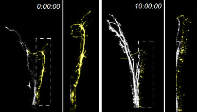 Selective degeneration of missorted axons (yellow)
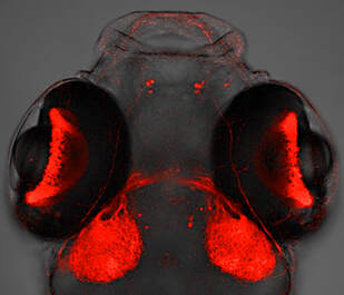
Retinal axons encounter many guidance decision points as they elongate towards their brain target, the optic tectum. Axons exit the eye through the optic nerve head, grow along the optic nerve toward the ventral midline of the diencephalon, cross the midline to form the optic chiasm, then navigate dorsally through the optic tract to reach the tectum where they establish connections with post-synaptic neurons. During this long navigation, some axons make mistakes at specific guidance points and take erroneous paths. They are however eliminated before reaching their final destination for proper wiring of the visual system. One axis of our research aims to dissect the mechanisms controlling the selective degeneration of misguided axons. In that context, we are particularly interested in determining the role of trans-axonal signaling in selective degeneration as well as the signaling pathways underlying the fragmentation and degeneration of axons.
Formation and maintenance of retinotopic maps
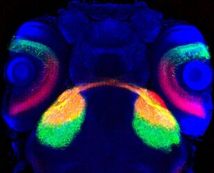
In the central nervous system, most axonal projections are organized into topographic maps according to the spatial organization of the neurons they originate from or the type of stimulus they respond to. In the visual system, retinal axons project topographically to the optic tectum according to their positional origin in the retina, thereby providing a co-herent representation of the external world. Axons originating from the more rostral retina project to the more posterior tectum (and conversely), and similarly, axons from the dorsal retina innervate the ventrolateral tectum (and conversely). A second axis of our research aims to characterize the molecular mechanisms controlling retinotopic map formation and maintenance. We are especially interested in determining the role of trans-axonal signaling and neuronal activity in map formation and refinement as well as the function of adhesion molecules in these processes.
